MEDICAL DEVICE AI BUILT IN TO READ RADIOLOGY
AI is rapidly changing the field of radiology, offering powerful tools to assist radiologists in analyzing medical images. Here’s how these “AI machines” work and their impact:
HOW AI READS RADIOLOGY IMAGES:
1. Image Acquisition: The process starts with acquiring medical images like X-rays, CT scans, MRIs, and ultrasounds.
2. Data Preprocessing: The images are preprocessed to enhance quality and standardize formats. This might involve noise reduction, image enhancement, and segmentation.
3. AI Algorithms: Sophisticated AI algorithms, often based on deep learning, are used to analyze the images. These algorithms are trained on vast datasets of labeled medical images to recognize patterns and identify abnormalities.
4. Feature Extraction: The AI system extracts relevant features from the images, such as the shape, size, texture, and location of organs, tissues, and potential abnormalities.
5. Analysis and Interpretation: The AI system analyzes the extracted features to identify potential abnormalities, classify them, and provide insights to the radiologist.
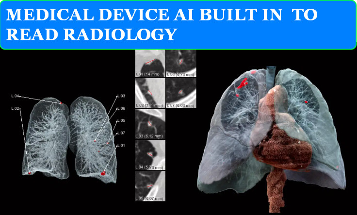
6. Output and Visualization: The AI system generates reports, highlights areas of concern on the images, and provides visualizations to aid the radiologist in interpretation.
EXAMPLES OF AI APPLICATIONS IN RADIOLOGY:
- Detection and Diagnosis:
- Aidoc: Flags acute abnormalities in CT scans, such as intracranial hemorrhages or pulmonary embolisms, to prioritize urgent cases.
- Viz.AI Contact: Analyzes CT scans to detect suspected strokes and alert specialists for faster treatment.
- Imagen OsteoDetect: Identifies distal radius fractures in wrist X-rays.
- Quantitative Analysis:
- AI-supported prostate segmentation and volume analysis: Helps measure prostate volume and identify suspicious lesions in MRI scans.
- Cardiac imaging analysis: Assesses cardiac function and detects abnormalities in echocardiograms or cardiac MRIs.
- Workflow Optimization:
- Prioritization of critical findings: Helps radiologists prioritize cases based on the urgency of the findings.
- Automated report generation: Assists in generating preliminary reports, saving radiologists time.
BENEFITS OF AI IN RADIOLOGY:
- Increased Accuracy: AI can detect subtle abnormalities that might be missed by the human eye, leading to more accurate diagnoses.
- Improved Efficiency: AI can automate tasks, allowing radiologists to focus on more complex cases and improving overall workflow.
- Reduced workload: AI can help alleviate the burden on radiologists, who often face heavy workloads and time constraints.
- Enhanced Patient Care: Faster and more accurate diagnoses can lead to earlier interventions and better patient outcomes.
IMPORTANT NOTE:
- AI as a Tool: AI is not meant to replace radiologists but to serve as a powerful tool to enhance their capabilities and improve patient care. The final interpretation and diagnosis always rest with the radiologist.
The use of AI in radiology is rapidly evolving, with new applications and advancements emerging constantly. This technology has the potential to revolutionize the field, making radiology more accurate, efficient, and accessible to patients worldwide.